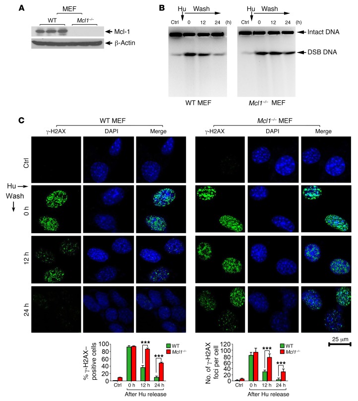
Figure 2. Knockout of Mcl-1 retards the repair of hydroxyurea-induced DSBs.
(A) Mcl-1 expression was analyzed by Western blot in WT and Mcl1–/– MEFs. (B and C) WT and Mcl1–/– MEFs were subjected to hydroxyurea (Hu, 0.2 mM) treatment for 24 hours. After Hu removal, cells were incubated in normal culture medium for various times up to 24 hours. Hu-induced DSBs were assessed by pulsed-field gel electrophoresis or immunofluorescence using γ-H2AX antibody at various time points. Scale bar: 25 μm. The percentage of γ-H2AX–positive cells (≥5 foci) and the number of γ-H2AX foci per cell were determined by counting of at least 100 cells from each sample. Data represent the mean ± SD, n = 3 per group. ***P < 0.001, by 2-tailed t test.

