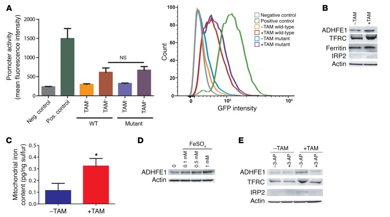
Figure 2. ADHFE1 promoter activity in HMEC-MYC cells and induction of ADHFE1 protein expression by FeSO4.
(A) HMEC-MYC cells were transfected with a reporter construct containing a 5-kb segment of the human ADHFE1 promoter region with a MYC binding site. Cells were transfected with constructs containing either the intact MYC binding site (WT) or a mutated sequence (mutant), as described in Methods. MYC signaling was induced with 4-hydroxytamoxifen (+TAM). Addition of TAM modestly increased ADHFE1 promoter activity but the activity was not dependent on the presence of an intact MYC binding site. Shown is the mean ± SD for triplicate experiments. Two-sided t test; NS, not significantly different. (B) MYC signaling (+TAM) increases expression of iron metabolism genes in HMEC-MYC cells. (C) MYC signaling also increases mitochondrial iron content in these cells. n = 4. Shown is the mean ± SD. *P < 0.05 (2-sided t test). (D) Treatment of HMECs with FeSO4 for 24 hours increases ADHFE1 expression. (E) Induction of ADHFE1 and iron metabolism genes by MYC signaling in HMEC-MYC cells can be inhibited by the Fe2+ chelator, 3-AP (0.5 μM), which was added together with TAM. In B, C, and E, TAM treatment was for 96 hours. TFRC, transferrin receptor protein 1; IRP2, iron-responsive element–binding protein 2.

