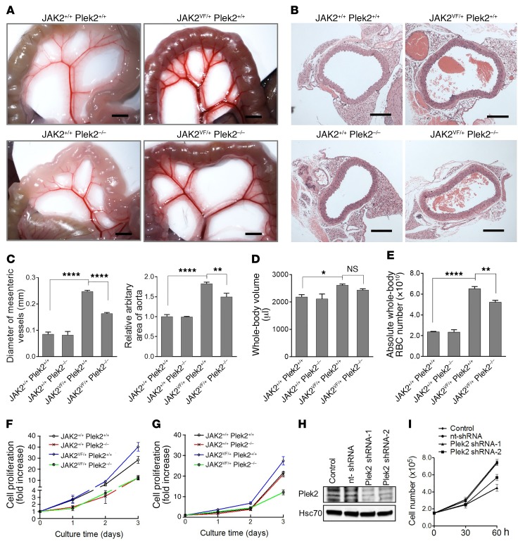
Figure 6. Reduction of RBC mass in JAK2V617F-knockin mice with the loss of Plek2.
(A) Representative mesenteric vessels at the same anatomic sites from the indicated mice. Scale bars: 2 mm. (B) Aorta cross section from the indicated mice. Scale bars: 200 μm. (C) Quantification of the diameters of the mesenteric vessels in A and aortic areas in B. N = 5 in each group. (D) Whole-body blood volume of the indicated mice. N = 5 in each group. (E) Calculated absolute circulating RBC (whole-blood volume × RBC count) of the indicated mice. N = 5 in each group. (F) Cell proliferation analyses from bone marrow lineage-negative cells of the indicated mice cultured in Epo-containing medium. Cells were counted using a hemocytometer. Data were obtained from 4 mice in each group. (G) Same as C except the cells were cultured in GM-CSF–containing medium. (H) Western blot assay to test the knockdown efficiency of shRNA Plek2 in SET-2 cells. Hsc70 is a loading control. (I) Cell proliferation assay of cells from H in the presence of Epo. Data were obtained from 3 independent experiments. *P < 0.05, **P < 0.01, and ****P < 0.0001; all P values were determined by 1-way ANOVA with Tukey’s multiple comparisons test.

