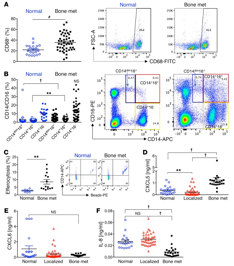
Figure 12. Nonclassical (CD16+) peripheral blood mononuclear cells and CXCL5 serum levels are associated with human prostate cancer skeletal metastasis.
(A–C) Mononuclear cells were isolated from whole peripheral blood of noncancer (Normal, n = 21) and prostate cancer bone-metastatic patients (Bone met., n = 47). Monocyte populations were assessed via flow cytometry. (A) CD68+ monocytes were assessed, and representative FACS images are displayed at right. (B) Subpopulations of CD14dimCD16+ (blue boxes), CD14+CD16+ (red boxes), and CD14+CD16– (yellow boxes) were gated from total cell populations and quantified for Normal and Bone met. samples. Representative FACS plots are shown. (C) Freshly isolated monocytes (CD14+) from normal (n = 8) and prostate cancer bone-metastatic (n = 14) patients were cultured with phosphatidylserine-coated fluorescently labeled apoptotic-mimicry beads (3:1) and efferocytosis assessed by flow cytometry for CD14+ (APC+) cells with ingested beads (representative FACS plots are shown). (D–F) Human serum isolated from normal (n = 20), localized (high-risk) prostate cancer (Localized, n = 40), and bone-metastatic prostate cancer (Bone met., n = 22) patients was analyzed by ELISA for CXCL5 (D), CXCL6 (E), and IL-8 (F). CXCL6 and IL-8 analysis included n = 38 for Localized. Data are mean ± SEM; **P < 0.01, #P < 0.001, †P < 0.0001 (Wilcoxon 2-sample test and Kruskal-Wallis test with Bonferroni’s correction).

