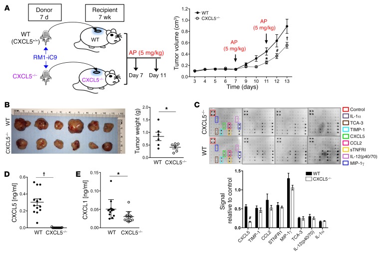
Figure 6. Prostate cancer growth in bone vossicles is hindered in CXCL5–/– mice.
(A) WT (CXCL5+/+) and CXCL5–/– recipient mice (7wk males) were implanted subcutaneously with RM1-iC9 inoculated vossicles isolated from WT or CXCL5–/– donor mice (7d males), respectively. Recipient mice from both groups were treated with AP at days 7 and 11 to induce apoptosis. Tumor volumes were quantified. An independent experiment with similar results is shown in Supplemental Figure 4A. Data are mean ± SEM; *P < 0.05, #P < 0.001, †P < 0.0001 vs. WT controls (2-way ANOVA). (B) Gross image of tumors and graph of quantified tumor weights; n = 6 per group. (C) Total protein lysates of tumor vossicles from WT and CXCL5–/– mice were analyzed via inflammatory cytokine array. Quantification of cytokines expressed is represented as signal relative to the positive controls in the array; n = 4 independent arrays per group. (D and E) Protein lysates were analyzed by ELISA for the expression of CXCL5 (D) and CXCL1 (E) in the tumor vossicles of WT and CXCL5–/– mice; n = 13 per group. Data in B–E are mean ± SEM; *P < 0.05, #P < 0.001, †P < 0.0001 (2-tailed Student’s t test).

