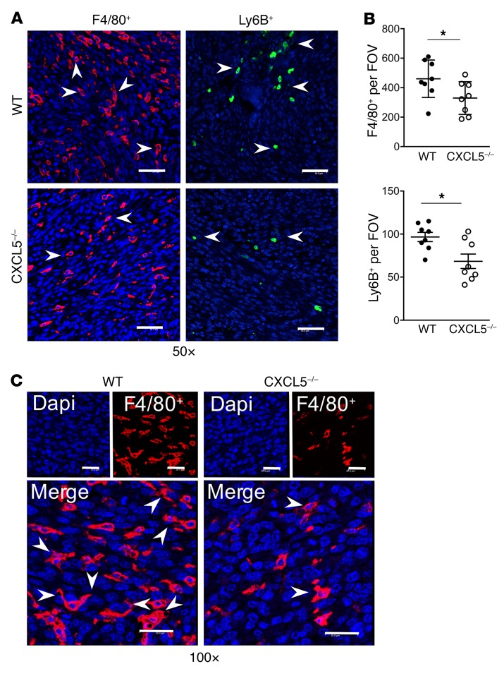
Figure 7. F4/80+ and Ly6B+ infiltration into tumor vossicles is hindered in CXCL5–/– mice.
(A) Representative fluorescence images of F4/80+ (Opal 570) and Ly6B+ (Opal 520) cells in tumor vossicle sections from WT and CXCL5–/– mice at ×50 original magnification (scale bars: 50 μm). (B) Quantification of F4/80+ and Ly6B+ staining (4 fields at ×10 per tumor vossicle sample); n = 8 per group. (C) Macrophage morphology at ×100 original magnification (scale bars: 30 μm) showing some MΦs with extended arms (engulfing-like shape) surrounding large nuclei (blue, DAPI) typical of cancer cells. Image depicts the WT and CXCL5–/– MΦs (red) infiltrating the tumor. Data in B are mean ± SEM; *P < 0.05 (2-tailed Student’s t test).

