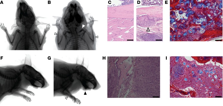
Figure 8. β-Catenin stabilization in bone progenitors promotes proliferation and blocks differentiation.
(A) Posteroanterior radiographs of a control mouse and (B) a Prx1CreERT2 Ctnnb1ex3fl mouse at 12 months of age show thickening of the clavicle (white arrowhead) and periosteal thickening (open black arrowheads) in the latter. (C) H&E histology photomicrographs of the ulnar cortex from a control mouse and (D) a Prx1CreERT2 Ctnnb1ex3fl mouse show the thickened periosteal mesenchyme layer, which contained (E) both osseous (red) and cartilaginous (blue) matrix production on Masson’s trichrome staining. Open arrowhead indicates the thickened periosteum. (F and G) Lateral projection radiographs of a control mouse and a Prx1CreERT2 Ctnnb1ex3fl mouse show the same forearm periosteal reaction (open arrowhead) as well as a bone matrix–forming mass arising from the jaw (black arrowhead) in the latter. (H) H&E-stained photomicrograph of one of these jaw masses shows a bland fibroblastic proliferation containing (I) islands of osseous (red) and cartilaginous (blue) matrix on the Masson’s trichrome–stained image. Scale bars: 100 μm.

