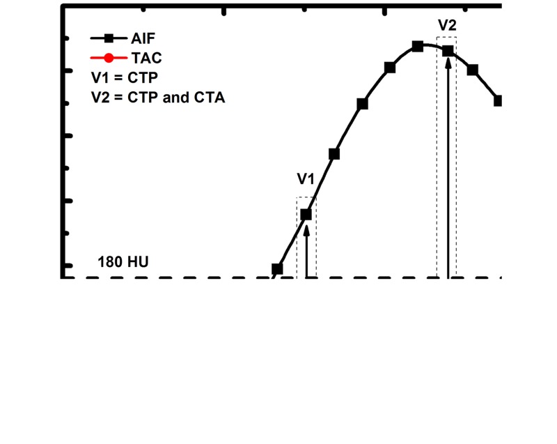
Figure 2:
To emulate a low-dose prospective acquisition protocol, two first-pass volume scans ( V1 and V2) are used for FPA perfusion measurement. The integrated change in myocardial attenuation (dMC/dt) is derived from the tissue time-attenuation curve ( TAC), and the average input concentration is estimated from the AIF. Both V1 and V2 are used for dynamic CT perfusion (CTP), while the volume scan after maximal attenuation ( V2) is also used for CT angiography (CTA).

