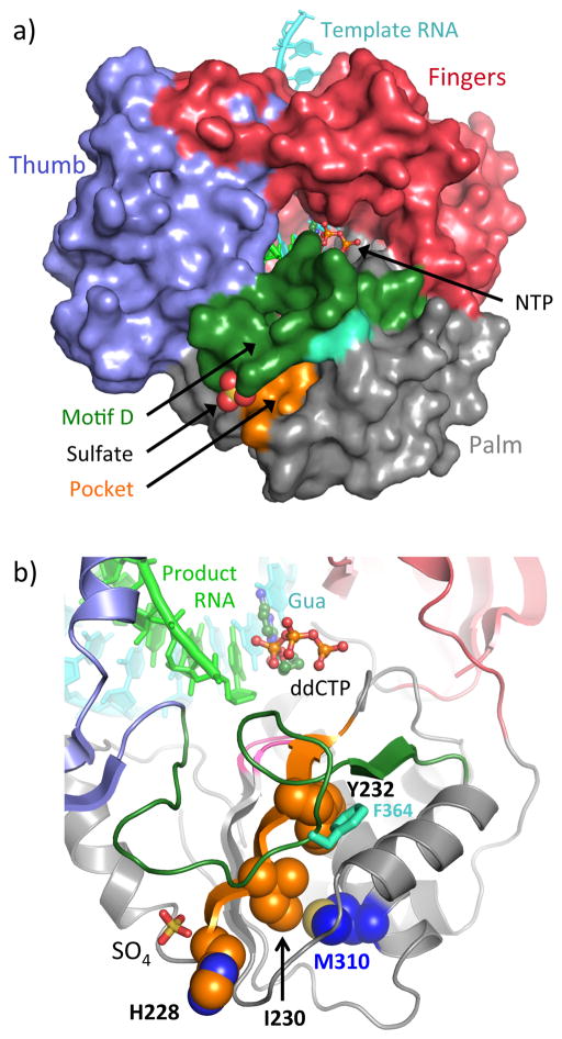
Figure 5.
Putative regulator binding pocket on CVB3 3Dpol based on loss of allosteric regulation by ATP in H228A/E, I230F, and F232Y mutants. A) Structure of the stalled CVB3 3Dpol elongation complex (PDB:4K4Y) with a bound sulfate from the apo-3Dpol structure (PDB:3DDK). Motif A residues lining the putative allosteric pocket are colored orange, motif D is colored dark green, and Phe364 is marked in lighter green. B) Detailed view of the mutated residues and their location relative to the active site with a bound dideoxy-CTP. Mutated motif A residues are shown as orange spheres and Phe364 in motif D is shown in light green sticks.

