
Figure 1.
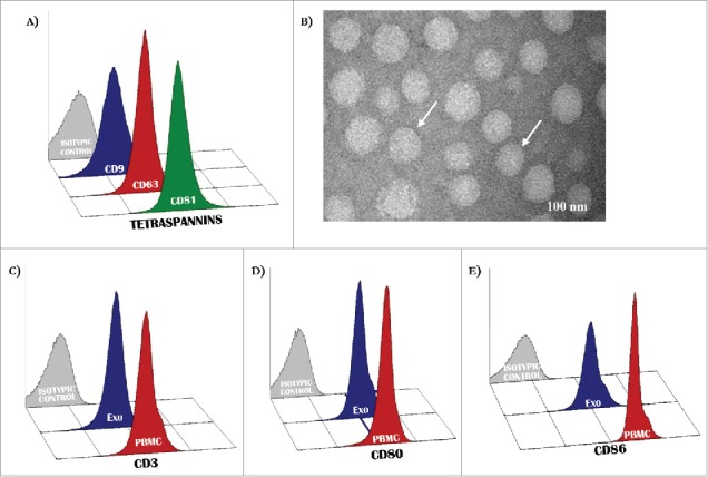
Antigenic profile of exosomes. (A) Exosomes isolated from patients with MM were characterized by flow-cytometry for the expression of CD9, CD63 and CD81 resulting in most instances higher than 95%. (B) Representative panels by TEM showing cup-shaped Exo (arrows) with a mean size of 80 nm (range: 40–110 nm). (C-D) A similar level of expression (higher than 90% in all instances) of CD3 as well as CD80 and CD86 antigens was revealed in peripheral mononuclear cells (red histograms) and Exo (blue histograms) isolated from 10 randomly selected MM patients. Histograms are representative of a single MM patient (pt.#3). Grey histograms are IgG1 a isotypic controls.