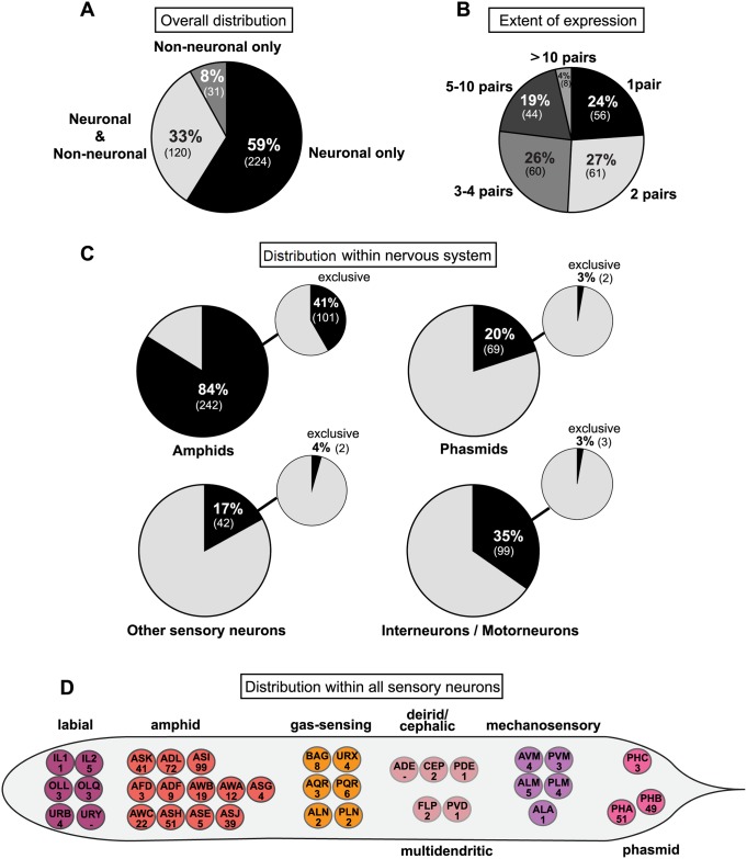
Fig 1. Summary of csGPCR reporter expression patterns.
(A) Overall tissue distribution of reporter expression patterns in hermaphrodites. Pie chart showing percentage of GPCR reporters expressed exclusively in neurons, in neurons and other cells types, and exclusively in non-neuronal tissues. Numbers in parentheses represent the absolute number of reporters in each category. (B) Extent of reporter expression within the nervous system. Pie chart showing percentage of neuronal reporters expressed in 1 neuron pair, 2 pairs, 3–4 pairs, 5–10 pairs, or more than 10 pairs. Numbers in parentheses represent the absolute number of reporters in each category. (C) Distribution of reporter gene expression within the nervous system. Pie charts showing percentage of GPCR reporters expressed in amphid neurons, phasmid neurons, other sensory neurons, and inter- or motorneurons. Small pie charts on the upper right represent the percentage of reporters exclusively expressed in amphids, phasmids, other sensory neurons, and inter- or motorneurons. Numbers in parentheses show the absolute number of reporters in each category. (D) Distribution within all sensory neurons of the hermaphrodite. Worm schematics showing the absolute number of GPCR reporters found to be expressed in each sensory neuron class. PHC is a phasmid neuron by name only. See S2 Table for a list of GPCR gene names expressed in the sensory neurons shown here. GPCR, G-protein-coupled receptor.

