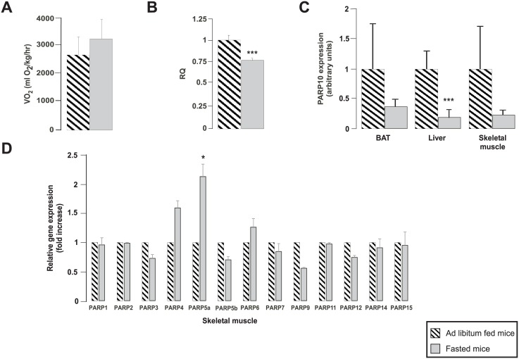
Fig 7. PARP10 expression changes inversely as mitochondrial oxidative capacity in mice.
C57/Bl6 male mice were subjected to 16 hours of fasting or received ad libitum food (n = 4/4, 3 months of age, average ± SEM). (A) The oxygen consumption and (B) the RQ were determined in these animals in indirect calorymetry experiments. (C) After dissection, the expression of PARP10 mRNA was determined by RT-qPCR in the brown adipose tissue (BAT), liver and skeletal muscle. (D) The expression of the members of the PARP family was determined in skeletal muscle using RT-qPCR reactions. Data is represented as average ± SEM. *, *** indicate statistically significant difference between control and transfected cells at p<0.05 or p<0.001, respectively.

