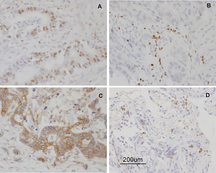
Fig 5. Immunohistochemical analysis of biopsied tumor specimens from patients.
(A) RNF43-positive cells in the tumor from patient KU-2. (B) Foxp3-positive lymphocytes in the tumor from patient KU-5. (C) HLA-Class1-positive cells in the tumor from patient KU-10. (D) CD8-positive lymphocytes in the tumor from patient KU-10 after treatment.

