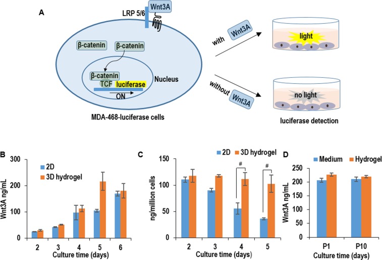
Fig 7. Quantitative detection of Wnt3A protein production in 2D adherent & 3D hydrogel cultures.
(A) MDA-468 cells (ATCC HTB-132TM) were stably transfected were with a luciferase reporter for the canonical Wnt signaling (Addgene, #24308). In the presence of biologically active Wnt3A proteins, the MDA-468-luciferase cells express luciferase proteins, which can be quantified with luciferase assay. (B) The concentration of Wnt3A proteins in the conditioned medium in 2D adherent & 3D hydrogel cultures from day 2 to day 6. (C) The protein productivity (i.e. ng of Wnt3A produced per cell per day) on day 2, 3, 4, and 5 in 2D adherent & 3D hydrogel cultures. (D) The Wnt3A concentration in the conditioned medium and hydrogel scaffold on day 5 in 3D hydrogel cultures. Error bars represent the standard deviation (n = 3). #: P<0.05.

