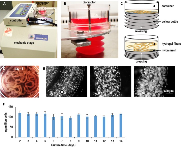
Fig 8. A prototype bioreactor for producing Wnt3A proteins.
(A, B, C) Single L-Wnt-3a-cells were mixed with 10% PNIPAAm-PEG solution and processed into hydrogel fibers with an extruder. Hydrogel fibers with cells were suspended within a column bioreactor. The medium was stored in a plastic bottle that could be pressed to flow the medium into or released to withdraw the medium from the bioreactor. (D) Image of hydrogel fibers with cells in the bioreactor. (E) Phase images of cells in hydrogel fibers on day 0, day 6 and day 14. (F) Cells consistently expressed Wnt3a during the two-week culture. Error bars represent the standard deviation (n = 3).

