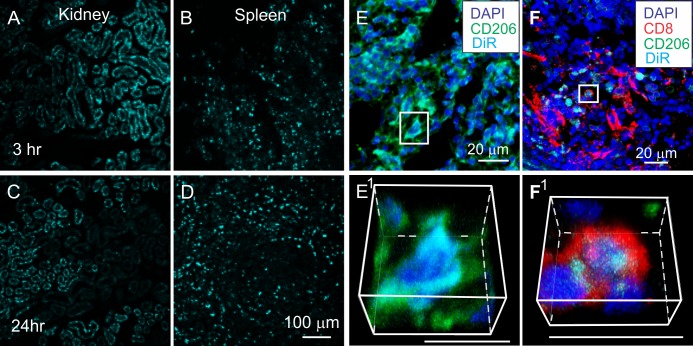
Fig 7. DiR hotspots transiently localize to kidney and more persistently to spleen.
A-D: DiR fluorescence (cyan) in kidney (A, C) and spleen (B, D) 3hours (A, B) and 24 hours (C, D) after infusion with DiR-MSCexos. Note the decrease in DiR fluorescence in the kidneys between 3 and 24 hours after DiR-MSCexos infusion, but increase in the spleen during the same time period. E-F: Spleens at 24 hours after DiR-MSCexos infusion in SCI rats immunostained with antibodies directed against CD206 (green) and CD8 (red) DiR fluorescence hotspots. E1-F1: Enlarged rotated images of boxed areas in E and F. Note that DiR hotspots localize to both CD206+ and CD4+ cells in the spleen. Scale bars in E 100 μm and applies to A-D. Scale bars in E&F indicate 20 μm.

