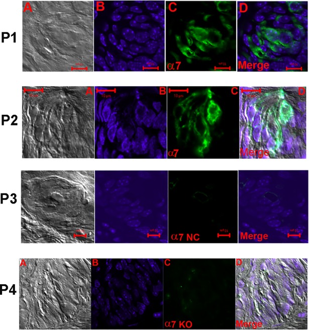
Fig 8. Immunostaining of α7 nAChR in WT mouse FF taste bud cells.
(A) DIC image (B) DAPI; (C) secondary antibody fluorescence (Alexa Fluor® 488; FITC) and (D) Merged image of DAPI and FITC. In panels (P1) and (P2) only a subset of TRCs within the FF taste buds showed binding to the nAChR α7 antibody. In panel (P2) nAChR α7 antibody binding was also observed in the apical pole of TRCs. In panels (P3) and (P4) no antibody binding was observed when the primary antibody step was omitted (NC) or in tissue sections from α7 KO mouse (KO), respectively. Horizontal bars = 10 μm.

