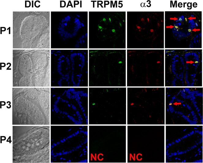
Fig 13. Immunostaining of α3 nAChR in enteroendocrine cells in the Trpm5-GFP mouse gut.
Shows DIC image, DAPI, Trpm5-GFP, α3 nAChR antibody binding (secondary Donkey/Rabbit-590 antibody), and the merged images of DAPI, GFP and 590 red-fluorescent dye. In each of the 8 slides from 3 different gut sections examined 1 to 4 enteroendocrine cells were positive for α3 nAChR in the crypts (P1, P2, and P3; red). The α3 nAChR-positive cells were also positive for Trpm5-GFP (green). Negative control (NC) without primary antibody (P4).

