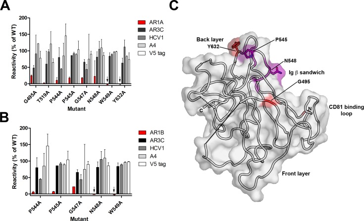
Fig 4. Epitopes for AR1 monoclonal antibodies target the non-neutralizing face of E2c.
Single residue mutations which resulted in ≤25% of mAb binding (relative to wild-type E2) but >75% for at least one control antibody are shown for mAbs AR1A (A) and AR1B (B). Binding assays were performed twice with the range indicated. Black arrows indicate negative values. (C) The critical residues for AR1A and AR1B were visualized on the E2c structure [15]. Residues in purple are required by both AR1A and AR1B. Residues specific for AR1A alone are in red. Dashed lines represent regions of E2c that are disordered or missing.

