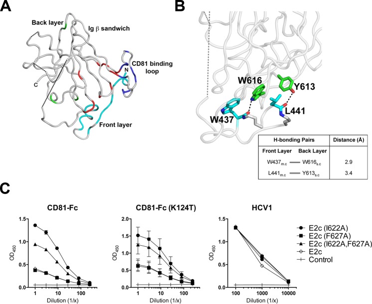
Fig 7. E2 back layer mutations modulate CD81 binding to the E2 front layer and CD81 binding loop.
(A) Based on flow cytometry and ELISA analysis, and previously published results [15], residues in four distinct E2c regions were found to be important for CD81 binding: the front layer (cyan), Ig β-sandwich (red), CD81 loop (blue), and the back layer (green). The locations of mutations resulting in <25% CD81 binding in each of these regions are highlighted. (B) Hydrogen bond calculations indicate that two back layer residues in the α2-helix, Y613 and W616 (green), interact with L441 and W437 (cyan) of the front layer α1-helix, respectively. m.c—main chain; s.c—side chain. These interactions between front and back layer residues suggest that the back layer indirectly affects CD81 binding through structural interactions with the adjacent front layer. The residue colors follows those in Fig 1C. (C) Based on flow cytometry and ELISA, I622A, which was found to enhance CD81 binding, was analyzed further (along with F627A). Soluble E2c mutants harboring I622A, I622A/F627A and F627A were tested using ELISA for their ability to bind recombinant WT CD81-Fc (left panel) or a mutant that reduces CD81 dimerization, CD81-Fc (K124T) (middle panel). Binding signals to mAb HCV1 were used as an expression control for the mutants (right panel).

