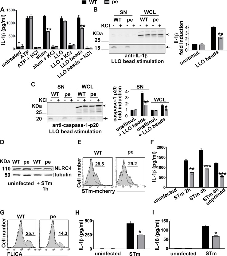
Fig 2. AP-3 promotes inflammasome activation triggered by phagocytosed stimuli.
A-C. BMDCs from WT or pearl (pe) mice were primed for 3 h with LPS and stimulated with the indicated NLRP3 inflammasome stimuli. After 4 h cell supernatants were collected and cell pellets were lysed and fractionated by SDS-PAGE. A. IL-1β was detected by ELISA on cell supernatants. B, C. pro-IL-1β and mature IL-1β (B) or caspase-1 and its p20 cleavage fragment (C) were detected by immunoblotting on both supernatants and cell lysates. Left, representative blots. Right, quantification of band intensities for mature IL-1β (B) or caspase-1 p20 (C) from three independent experiments, showing fold increase relative to unstimulated cells and normalized to whole cell lysates (mean ± SD). D-I. BMDCs (D-G) or splenic DCs (H, I) were uninfected or infected with flagellin-expressing STm (stimulates NLRC4) at 10 MOI. D. Representative immunoblot showing NLRC4 expression and tubulin as loading control. E. Uninfected cells (dotted lines) or cells infected for 15 min with mCherry-labeled STm (STm-mCherry; gray filled lines) were washed and analyzed by flow cytometry. Shown is a representative of 3 independent experiments. Numbers represent percent of cells with above background mCherry signal. F. At the indicated time points after infection with STm, cell supernatants were collected and assayed for IL-1β by ELISA. G. The caspase-1 probe FAM-FLICA was added after STm infection. After 15 min, cells were washed and analyzed by flow cytometry. Dotted lines, uninfected cells; gray filled lines, infected cells. Shown is a representative experiment. H, I. Supernatants from splenic DCs that were untreated or infected for 4 h with STm were collected and assayed for IL-1β (H) or IL-18 (I) by ELISA. Data in D-I are from three independent experiments; data in F, H and I show mean ± SD. *p<0.05; **p<0.01; ***p<0.001.

