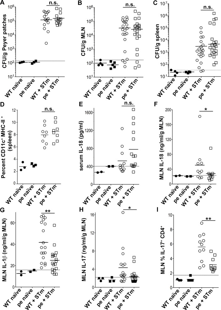
Fig 4. AP-3-deficient mice produce less IL-1β, IL-18 and IL-17 early after Salmonella Typhimurium infection.
WT and pearl (pe) mice were infected orally with 109 STm or received PBS as a control and analyzed three days after infection. (A-C). Peyer patches, MLN and spleens were harvested, homogenized and plated to measure bacterial load. CFUs were normalized to tissue weight (expressed as CFU/ g of tissue). D. Cell suspensions from spleen were analyzed by flow cytometry for CD11c+ MHC-II+ DCs. Average cell number: 250x106 E. Blood was collected by cardiac puncture, and serum was isolated and assayed for IL-18 by ELISA. (F-H). Supernatants from homogenized and pelleted MLN were assayed for IL-18 (F), IL-1β (G) and IL-17 (H) by ELISA. I. Single cell suspensions from MLN were restimulated in vitro for 5 h with 10 ng/ml PMA, 1 μg/ml ionomycin and 10 μg/ml of brefeldin A. Cells were then fixed, permeabilized, stained for intracellular IL-17 and analyzed by flow cytometry, gating on CD4+ T cells. Data in each panel are pooled from three independent experiments. Dotted lines, background (threshold values from uninfected mice). Solid lines, geometric mean (A-C) or arithmetic mean (D-I) of values from infected mice. *p<0.05; **p<0.01; n.s., not significant. See also S3and S4 Figs.

