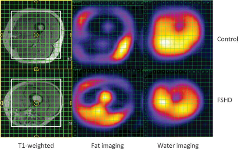
Fig. 7.

With multivoxel MR spectroscopy, 16 × 16 grids were placed over the midthigh of a healthy volunteer (top left) and a patient with facioscapulohumeral muscular dystrophy (FSHD) (bottom left). After Fourier transformation, corresponding spectra in the second and third columns show elevated fatty infiltration of the midthigh musculature in the FSHD patient, particularly in the posterior thigh in the region of the adductors and hamstrings. The control patient demonstrates a normal appearance and minimal degree of fatty infiltration of muscles of the midthigh.
