
Figure 5. M1 action as natively expressed on GAS in vitro and in vivo and as soluble protein in vivo.
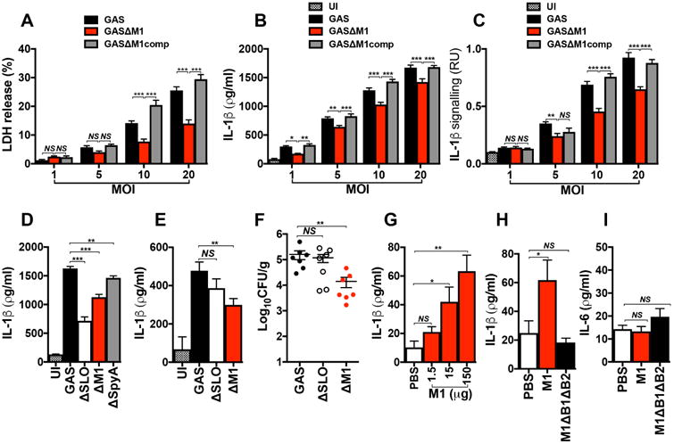
THP1-Mϕ were coincubated with WT (GAS), isogenic Δemm1 mutant (GASΔM1) or M1-complemented (GASΔM1comp) strains at different multiplicities of infection (MOI). After 2 h of infection, macrophage supernatants were collected and analyzed for detection of LDH (a), total IL-1β (b) and mature IL-1β (c). d, Comparison of IL-1β secretion from THP1-Mϕ coincubated for 2 h with WT GAS or isogenic Δemm1 (GASΔM1), Δslo (GASΔSLO) and ΔspyA (GASΔSpyA) mutant strains. Uninfected macrophages were used as negative control (UI). IL-1β production was analyzed by ELISA and the presence of mature IL-1β by HEK-Blue™ IL-1R reporter cells. IL-1β detection (e) and bacteria recovered (CFU) (f) from peritoneal lavage fluid of wild type C57BL/6 mice 6 h after intraperinoneal infection with 1 × 10⁸ CFU of WT or isogenic ΔM1 or ΔSLO mutant strains. Control group mice were injected with PBS and used as negative control (UI). Detection of IL-1β (g and h) and IL-6 (i) from peritoneal lavage fluid of WT C57BL/6 mice, 4 h after injections with different concentrations of M1 protein (g), and with 150 μg of purified M1 or ΔB1ΔB2 proteins (h and i). Control group animals were injected with PBS. Cytokine quantification was performed by ELISA. Data in panels a-i are plotted as the mean ± SEM. Panels a, b, c, and d represent three independent experiments performed in triplicate. In panels e and f N=7, in panel g N=6 and in panels h and i N=10 and represent the combination of 2 independent experiments. Results in panels a-c were analyzed by Two-way ANOVA multiple comparisons and panels d-i were analyzed by Student's t-test. NS = not significant (P>0.05), * P<0.05, ** P<0.01 and ***P<0.001).