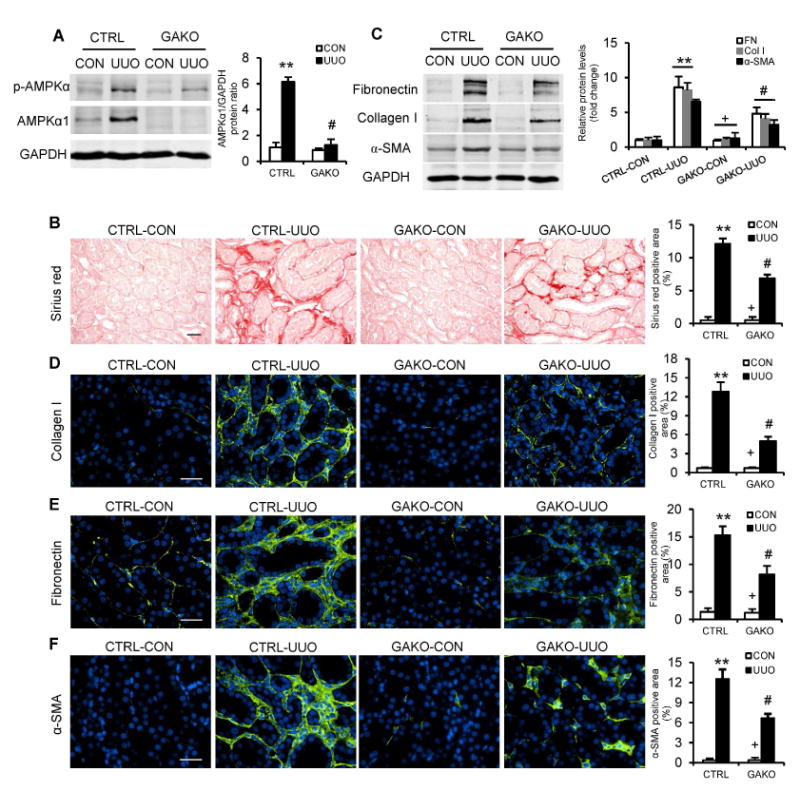
Fig. 1. Global AMPKαl deficiency attenuates renal fibrosis induced by UUO.

(A) Representative Western blots show the protein levels of AMPKαl and p-AMPKα in the contralateral (CON) and UUO kidneys of GAKO mice and their littermate controls (CTRL). Protein expression was normalized with GADPH. (B) Representative photomicrographs show kidney sections stained with Sirius red for total collagen deposition. Scale bar: 20 μm. The bar graph shows quantitative analysis of renal interstitial collagen content in different groups. (C) Representative Western blots show protein levels of fibronectin (FN), collagen I (Col I), and α-smooth muscle actin (α-SMA) in the kidneys. Representative photomicrographs of the kidney sections stained for collagen I (D), fibronectin (E) and α-SMA (F), and counterstained with DAPI (blue). Scale bar: 30 μm. Bar graphs show quantitative analysis of protein immunostaining in the kidney sections. **P<0.01 compared with the CTRL-CON group, #P<0.05 compared with the CTRL-UUO group, +P<0.05 compared with the GAKO-UUO group. n=6 mice per group.
