
Figure 2. RAPTOR structure and TOS motif recognition.
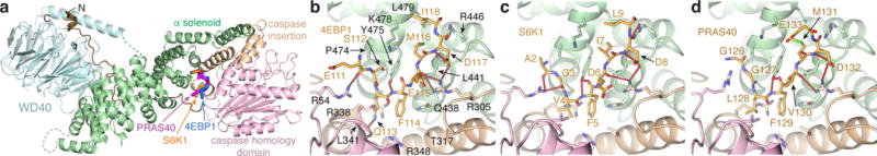
a, atRaptor structure with the three TOS motif co-crystal structures superimposed. atRaptor domains colored as labelled (N-terminal extension dark brown). b–c, Close-up views of the interface between atRaptor, colored as in a, and TOS peptides (yellow sticks) of human 4EBP1 (b), S6K1 (c) and PRAS40 (d). atRaptor side chains shown are identical in human RAPTOR, whose residue numbers are shown (red-dotted lines, hydrogen bonds).