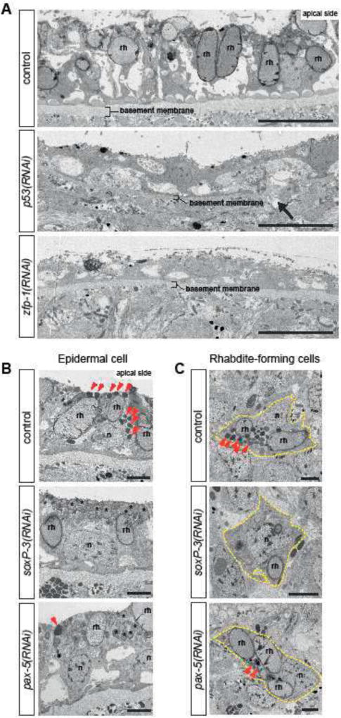
Figure 4. Ultrastructural analysis show distinct epidermal defects inp53-, zfp-1-, soxP-3- and pax-2/5/8 (RNAi) animals.
(A) TEM micrographs of ventral epidermis. p53-and zfp-1(RNAi) epidermis appear to be stretched, contain reduced number of rhabdites (rh) and sit on thinner basement membrane (arrow points to a rupture). n = 3, 100% penetrance. Scale bar, 10µm. (B) TEM micrographs of dorsal epidermis. The numbers of electron-dense granules (arrowheads) in the epidermal cells are greatly reduced by soxP-3-and pax-2/5/8 RNAi. Instead, the apical side of soxP-3 and pax-2/5/8(RNAi) epidermis are filled with rhabdite-like vesicles (asterisks). n, nucleus; rh, rhabdite. Scale bar, 2µm. (C) TEM micrographs of rhabdite-forming cells. Electron-dense granules (arrowheads) observed in the control cell are greatly reduced in soxP-3-and pax-2/5/8(RNAi) cells. Scale bar, 2µm.

