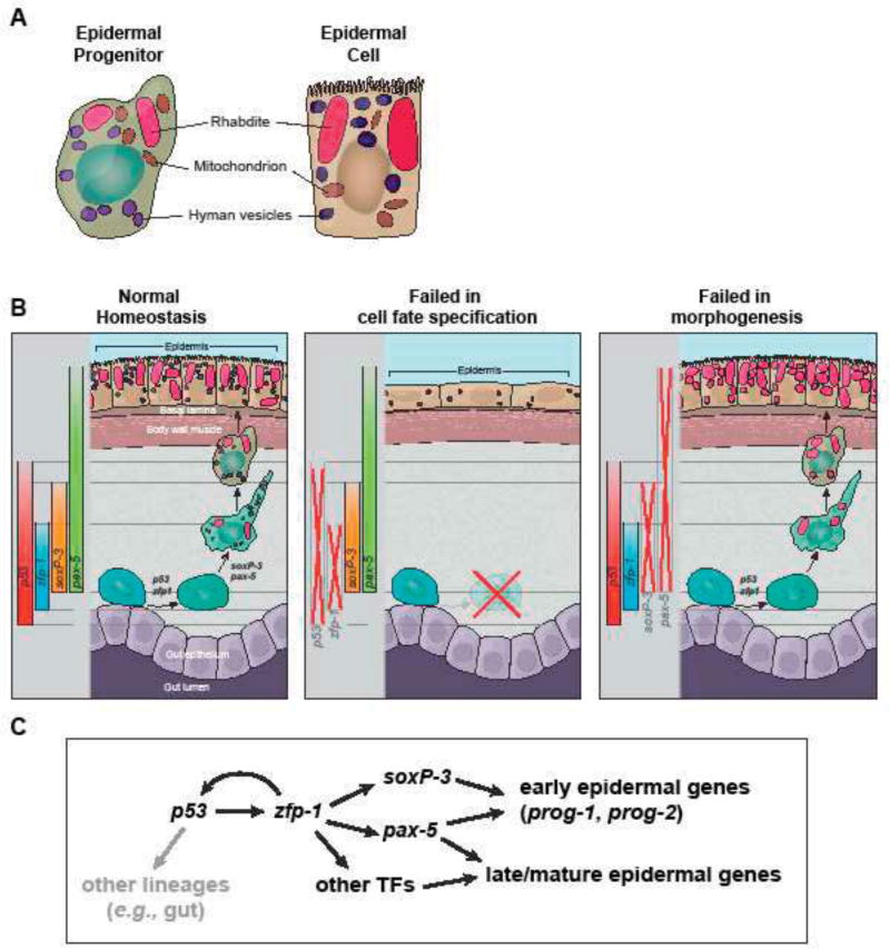
Figure 7. Model of epidermal lineage progression in planarian.
(A) Schematic of ultrastructures of epidermal precursor and mature epidermal cell. Epidermal precursors contain features of the mature epidermal cells including large rod-shape rhabdites and small oval-shape granules. They are also abundant in mitochondria. (B) Schematic of the planarian epidermal lineage progression and the phenotypic differences in p53-, zfp-1-, soxP-3 and pax-2/5/8 (RNAi) animals. (C) Proposed transcription network in the epidermal lineage progression. Gray arrow represents aspects of the network not studied in depth.

