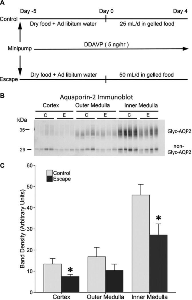
Figure 1. Experimental model.

A. The study design was adapted from Ecelbarger et al.3. Rats were euthanized on day 1, 2, and 4. B. Immunoblotting for AQP2 in the cortex, outer medulla, and inner medulla (n=4 per group) at day 2 of escape protocol. C. Densitometry shows a significant decrease in aquaporin-2 protein abundance in the cortex and inner medulla of the escape animals, consistent with previous reports. For each lane, 10 g (cortex), 5 μg (outer medulla), and 2 μg (inner medulla) of total protein was loaded. *p < 0.05 by unpaired t-test. (Coomassie-stained gels, run in parallel, showed equal loading.) Glyc-AQP2, glycosylated aquaporin-2; non-Glyc-AQP2, nonglycosylated aquaporin-2.
