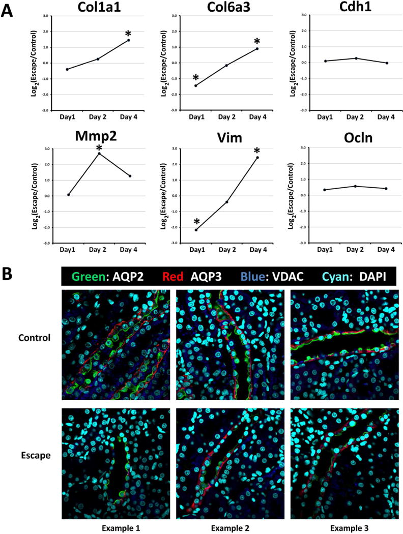
Figure 6. Onset of vasopressin-escape is associated with a partial epithelial-to-mesenchymal transition in rat CCD cells.

A. Time course of transcript abundance changes for selected EMT marker genes during onset of vasopressin-escape in microdissected rat CCDs shows increase in mesenchyme-associated transcripts (Col1a1, Col6a3, Mmp2 and Vim) without loss of epithelium-associated transcripts (Cdh1 and Ocln). Asterisk indicates Benjamini-Hochberg FDR-adjusted P value <0.05 (see Supplementary Table 2 for standard errors). B. Immunocytochemical labeling for AQP2 and AQP3 in renal cortex of rats undergoing vasopressin-escape shows retention of normal epithelial polarity. VDAC labeling of mitochondria was also carried out to reveal presence of intercalated (‘mitochondria-rich’) cells. DAPI labeling of nuclei facilitates recognition of apical versus basal aspects of cells.
