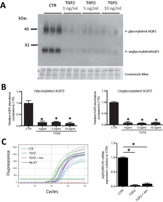
Figure 7. TGFβ exposure decreases AQP2 protein and mRNA abundance in mpkCCD cells.

A. Representative immunoblot showing relative AQP2 abundances in control (CTR) cells and cells exposed to TGFβ (1, 5 or 10 ng/ml) for 2 days in serum-free medium. The cells were pretreated with 1 nM dDAVP on the basolateral side for 4 days to induce AQP2 expression. Bottom panel shows a Coomassie-stained gel to demonstrate equal loading. B. AQP2 band density was significantly decreased both for glycosylated and nonglycosylated AQP2 (n=3). C. SYBR Green™ fluorescence curves for RT-qPCR experiments quantifying AQP2 mRNA under the control (CTR) condition, after 1 ng/ml TGFβ (2 days) and after 1 ng/ml TGFβ plus amiloride at 10 μM (2 days). Cells were pre-treated with dDAVP at 1 nM for 4 days. Horizontal green line is the threshold used to calculate Ct. The relative AQP2 abundances (normalized to the housekeeping gene RPLP0) are shown as a bar graph.
