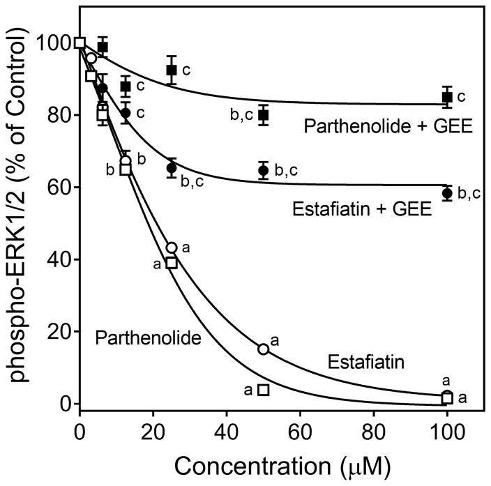
Figure 4.
Effect of parthenolide and estafiatin on activation-induced ERK1/2 phosphorylation. Jurkat T cells were pretreated with 1% DMSO or increasing concentrations of estafiatin (○) or parthenolide (□) for 20 min, followed by activation with anti-CD3/CD28 (10 μg/ml each) for 5 min, and the levels of ERK1/2 phosphorylation were evaluated using ELISA. In some experiments, Jurkat cells were incubated overnight with 2 mM GEE or medium (not shown), followed by treatment with DMSO or increasing concentrations of estafiatin (●) or parthenolide (■) for 20 min, followed by activation with anti-CD3/CD28 (10 μg/ml each) for 5 min, and ERK1/2 phophorylation was evaluated using the same procedures described above. The data are presented as % of control levels and represent the mean ± SD of triplicate samples from one experiment, which is representative of three independent experiments. Statistically significant differences between cells pretreated with sesquiterpene lactones versus control (1% DMSO) are indicated (ap<0.01; bp<0.05). In addition, statistically significant differences between cells incubated with GEE versus without GEE prior to the respective lactone treatment and activation are indicated (cp<0.01).

