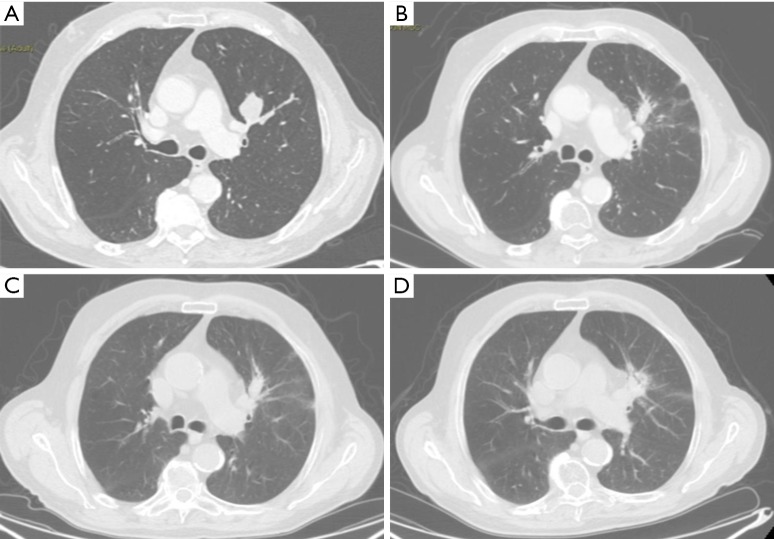
Figure 2.
CT images of benign and high-risk CT features. (A) Eighty-three years old male, CT scan pre-SBRT, NSCLC stage IB; (B) 6 months after SBRT: scar-like fibrosis, patchy GGO; (C) 12 months after SBRT: no differences respect the first one; (D) 24 months after SBRT: mass-like fibrosis. CT, computed tomography; SBRT, stereotactic body radiation therapy; NSCLC, non-small cell lung cancer; GGO, ground glass opacities.

