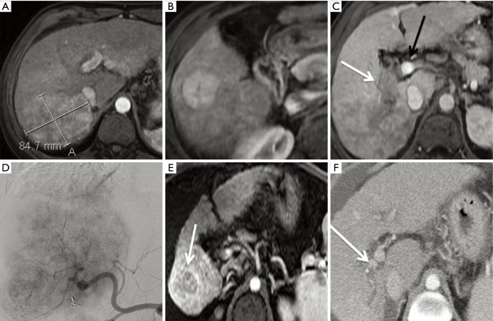
Figure 5.
Y-90 SIRT for BCLC C hepatocellular carcinoma. This 60-year-old female with chronic hepatitis C was imaged after she had elevated liver function tests during a routine physical examination. Magnetic resonance imaging demonstrated an 8.7 cm right posterior (A) and smaller satellite nodule more inferiorly (B). The right portal vein was thrombosed and distended with tumor (white arrow, C), while the main and left portal veins remained patent (black arrow, C). At a multidisciplinary tumor board, the group decision was made to treat with intra-arterial therapy and to reserve sorafenib for metastatic disease should it develop at a later time. Angiography demonstrated the extensive tumor occupying most of the right lobe (D). Two months after treatment, there was remarkable reduction in tumor size and enhancement burden, with a small residual inferior nodule most notable (arrow, E). Three months later, the tumor thrombus had also decreased in size and the right portal vein had partially recanalized (arrow, F). Y-90, yttrium 90; SIRT, selective internal radiation therapy; BCLC, Barcelona clinic liver cancer.

