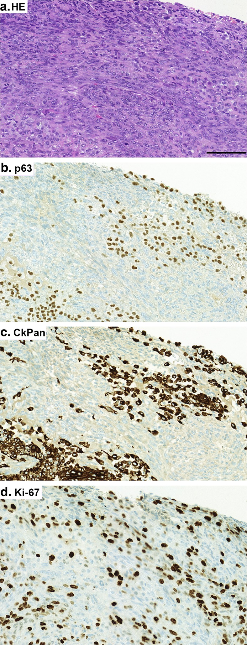
Fig. 3.
Representative images of selected immunohistochemical stainings of the massive breast tumour. HE staining (a). Immunohistochemistry (IHC) for p63 (b). IHC for CkPan (c). IHC for Ki-67 (d). Positive immunoreactivity can be seen in brown. Scale bar 100 μm. Note that there are areas within the tumour showing immunoreactivity for both CkPan and p63 (3b and c). This can be a display of carcinomatous epithelium transforming into metaplastic component

