Fig. 2.
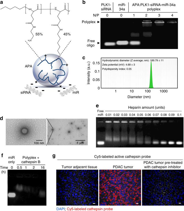
Physicochemical characterization of APA-miRNA–siRNA polyplexes. a Schematic illustration of APA nanocarrier complexed with small RNAs (polyplex) and chemical structure of APA polymeric nanocarrier. b Polyplex formation of APA with miR-34a and PLK1-siRNA (total of 50 pmol oligonucleotides, miRNA/siRNA ratio of 1:1) showing the optimal Nitrogen/Phosphate (N/P) ratio using EMSA. c Hydrodynamic diameter and surface charge of the polyplex at N/P ratio of 2, measured by particle size analyzer and Zetasizer, respectively. d Representative TEM images of the polyplex. e miR-34a release from the polyplex obtained in vitro by the polyanion heparin displacement assay. f miR-34a release from the polyplex by cathepsin B (2 units per mg polymer) cleavage of the PGA backbone. g Direct labeling of active cathespins in PDAC tumor xenograft and in normal adjacent tissues. Frozen sections were fixed on slides, incubated with 0.25 µM Cy5-labeled cathepsin activity-based probe (in red), stained with 4′,6-diamidino-2-phenylindole (DAPI, in blue) and imaged with fluorescent microscope. For specificity of staining, additional slides were treated with a non-labeled cathepsin inhibitor (GB111, 5 µM) prior to incubation with the Cy5-labeled cathepsin activity-based probe (right image). Scale bar, 10 µm

