Fig. 1.
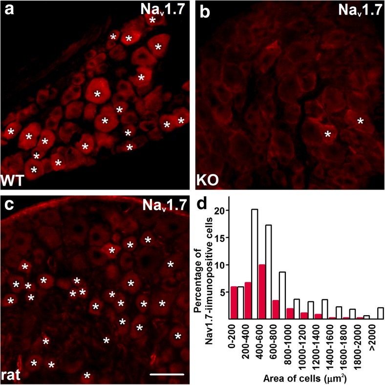
The anti-Nav1.7 antibody specifically identifies a sub-population of primary sensory neurons. a Incubation with the anti-Nav1.7 antibody (Millipore, AB5390) results in immunostaining in a group of primary sensory neurons in wild-type (WT) mice. Asterisks indicate immunopositive neurons. b The same anti-Nav1.7 antibody produced only faint staining in very few neurons in sections cut from the dorsal root ganglia dissected from mice lacking Nav1.7 (KO; asterisks). c The anti-Nav1.7 antibody also produces immunostaining in rat primary sensory neurons. Immunopositive cells are indicated by asterisks. d Cell size distribution of Nav1.7+ neurons in the L4 and L5 dorsal root ganglia of naive Sprague-Dawley rats. Note that most of the Nav1.7+ neurons are small- and middle-size cells. Empty bars indicate size distribution of all cells whereas red bars indicate the size distribution of neurons exhibiting Nav1.7 immunopositivity. Though we have not tested the expression of functional Nav1.7, the specificity and selectivity of the antibody suggest the expression of such functional channels. Scale bar = 50 μm on each microphotographs (Color figure online)

