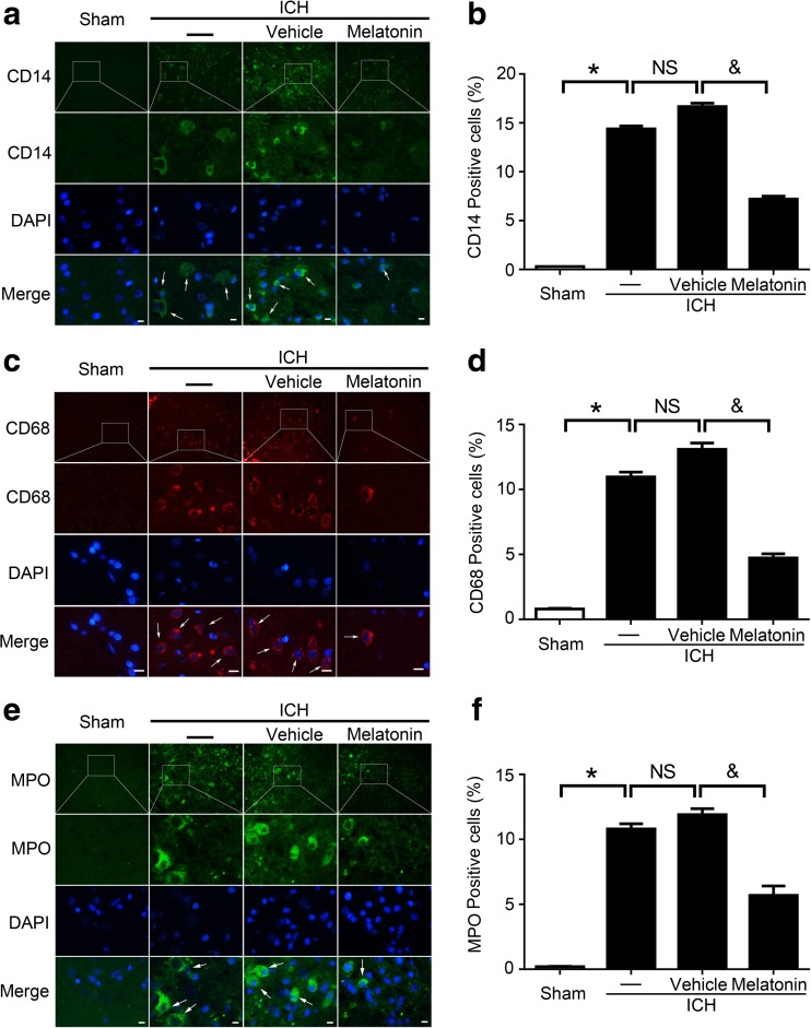
Fig. 5.
Immunofluorescence (IF) staining for the identification of inflammatory cells around the hematoma in brain tissues at 72 h post-ICH. Representative IF staining to identify CD14-positive (a), CD68-positive (c), and MPO-positive (e) cells (green or red), with the nuclei fluorescently labeled with 4,6-diamino-2-phenyl indole (DAPI, blue); scale bar = 32 μm. Percentage of CD14-positive (b), CD68-positive (d), and MPO-positive (f) cells around the hematoma in the brain tissues. Arrows indicate CD14-positive, CD68-positive, and MPO-positive cells. All data are displayed as mean ± SEM, with *P < 0.05 and & P < 0.05 deemed as significant difference; NS, no significant difference, n = 6

