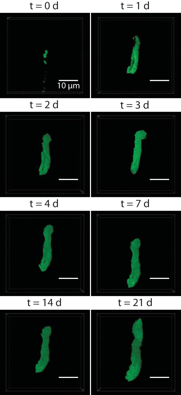
FIG 6 .

Confocal micrographs of a bacterial population in a receptacle over the course of 3 weeks. An individual living immature IJ nematode (head toward the top) was maintained in the microfluidic device, and the GFP-expressing bacterial population in the receptacle was imaged on days 0, 1, 2, 3, 4, 7, 14, and 21 posttrapping. Bacterial populations were sectioned in 300-nm z-steps.
