
Figure 6.
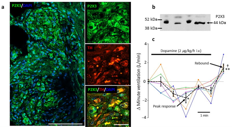
P2X3 receptor expression and aberrant tone generation in hypertensive human carotid bodies. (a), on the left is a section of a carotid body from a human cadaver showing P2X3 receptor immunofluorescence (green) with the nuclear stain - DAPI (blue; scale bar 100 μM). On the right, high power confocal images showing co-localisation of P2X3 receptors, tyrosine hydroxylase (TH) and DAPI (scale bar 25 μM). Data repeated in five human carotid bodies. See Supplementary Fig. 10 for P2X3 receptor antibody control and Supplementary Fig. 11 for axonal labelling. (b), western blot of the P2X3 receptor protein in human carotid body (n = 4). (c), low dose dopamine infusion (2 μg/kg/h i.v.) was used to inactivate the carotid bodies in six awake hypertensive humans (each colour coded) while recording minute ventilation in the supine position. A dextrose vehicle infusion was used as control and reference at time zero. The mean (± s.e.m.) response is indicated by the black dotted line. Note appearance of a rebound hyperventilatory response post-dopamine infusion. One-way ANOVA Bonferroni post-test. * P < 0.05 vehicle versus peak depression; ** P < 0.01, depression versus rebound (mean of 5 min); † P < 0.05, vehicle versus rebound.