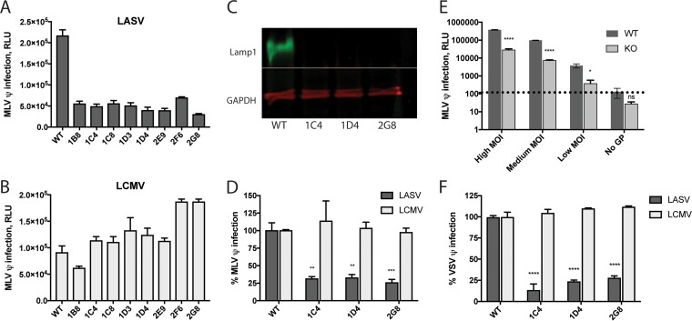
FIG 2 .
Knockout of Lamp1 reduces but does not abolish LASV GPC-mediated infection. Eight Lamp1 KO clones (see Materials and Methods) were screened for infection with LASV (A) and LCMV (B) MLV pseudoviruses. Data represent average luminescence units ± SD, measured in triplicate, from one experiment. (Two additional experiments with similar results were performed.) The level of LASV infection across these eight Lamp1 KO clones was 22% ± 5.3% of that seen in WT cells. (C) Representative blot (out of five similar blots) of lysates from three KO clones showing that Lamp1 was not detectable. (D) The above clonal KO lines were infected in quadruplicate with a moderate input (titer determined for a signal of ~100,000 relative light units [RLU] in WT cells) of either LASV or LCMV MLV luciferase pseudoviruses. Data were normalized to maximal signal from WT cells, and statistical significance was calculated by comparing the percentage of infection in KO cells against the percentage of infection in WT cells using a two-way analysis of variance (ANOVA). Error bars represent SD. **, P < 0.01; ***, P < 0.001. (E) One representative clone (2G8) was assayed in triplicate for infection with high, medium, and low input levels of LASV GPC pseudoviruses. Pseudoviruses lacking glycoprotein (“No GP”) were used to establish a background signal, indicated by a dashed line. Error bars represent SD. *, P < 0.05, ****, P < 0.0001, and ns, not significant, based on multiple unpaired, two-tailed t tests. The experiment was repeated twice with similar results. (F) The same clonal KO lines tested in panel D were infected with LASV and LCMV VSV pseudoviruses encoding GFP, and the titer was determined for 40 to 70% infection in WT cells. Signal from the “No GP” control pseudoviruses was subtracted from all measurements. As in panel D, infection signal from triplicate samples was normalized to WT cells and statistical analysis was applied to the efficiency of infection in KO cells compared to WT cells. The experiment was repeated two times with similar results.

