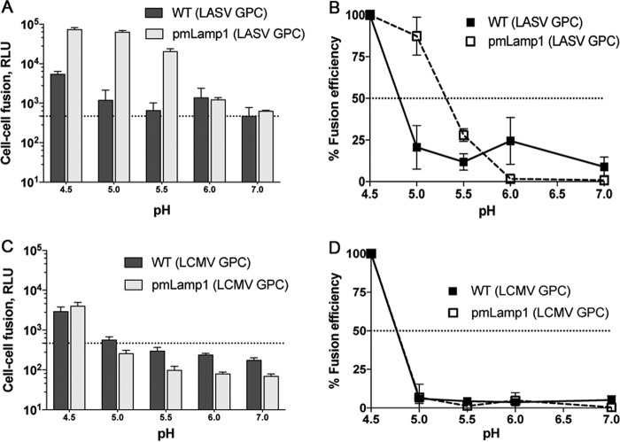
FIG 4 .
Lamp1 increases the extent and raises the pH threshold of LASV GPC-mediated fusion. In panels A and C, luminescence shows the extent of cell-cell fusion with WT (dark boxes) or pmLamp1 (light boxes) cells for LASV (A) and LCMV (C). Data represent RLU ± SD from the average of triplicate measurements. Dashed lines in panels A and C indicate background signal. In panels B and D, the data were normalized to fusion at pH 4.5 and replotted to show the corresponding pH dependence of cell-cell fusion for LASV (B) and LCMV (D). Dashed lines in panels B and D indicate 50% fusion efficiency. The LASV experiment was performed two additional times with similar results. Error bars represent the average ± SD of normalized values.

