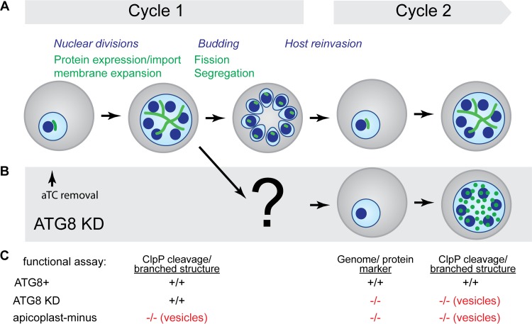
FIG 5 .
Schematic summarizing apicoplast defects detected in ATG8-deficient parasites. (A) Time course of cellular events during blood-stage development of Plasmodium parasites (the apicoplast is shown in green, the parasite nuclei and cell membrane are shown in blue, and the host erythrocyte is shown in gray). Steps in apicoplast development are indicated in green font, and those in parasite replication are indicated in italic blue font. (B) Apicoplast biogenesis defects observed upon ATG8 knockdown (KD) are shown. The timing of aTC removal to start ATG8 knockdown in ring-stage parasites is indicated (arrow). (C) Summary of functional assays performed in this study to assess the effects of ATG8 knockdown on specific steps in apicoplast development listed in panel A. The results of these functional assays are compared in ATG8+, ATG8 knockdown, and apicoplast-minus parasites, with defects highlighted in red font.

