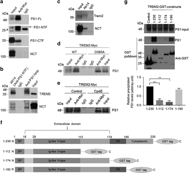
Figure 1.
Presenilin 1 (PS1) interacts with TREM2. (a, b) PS1 constructs were transfected into HEK293 cells stably expressing TREM2 with a Myc-tag at the C terminus (HEK293-TREM2). (a) Cell lysates were immunoprecipitated with a Myc antibody or control IgG. Immunoprecipitated proteins were subjected to immunoblotting with an Ab14 antibody to detect full-length PS1 (PS1-FL) and the PS1 N-terminal fragment (NTF), an anti-PS1 loop antibody to detect the PS1 C-terminal fragment (CTF) and a nicastrin (NCT) antibody as indicated. (b) Cell lysates were immunoprecipitated with Ab14, anti-PS1 loop or control IgG and immunoblotted with Myc and NCT antibodies. (c) Lysates from BV2 microglial cells were immunoprecipitated with Ab14 and immunoblotted with NCT and mouse TREM2 antibodies. (d) Vectors expressing wild-type (WT) or mutant PS1 (D385A) were transfected into HEK293-TREM2 cells. Cell lysates were immunoprecipitated with Ab14 or control IgG, and PS1 was detected by immunoblotting. (e) Lysates from HEK293-TREM2 cells with or without Compound E (CpdE, a γ-secretase inhibitor) treatment were immunoprecipitated with Ab14 or control IgG, and PS1 was detected by immunoblotting. (f) Schematic representations of full-length (1–230) or truncated TREM2 constructs, all tagged with GST at the C terminus. SP, signal peptide; TM, transmembrane domain. (g) PS1 was co-expressed with full-length TREM2 or other TREM2 fragments as shown in f in HEK293 cells. Cell lysates were precipitated with Glutathione Sepharose beads and immunoblotted with the PS1 antibody Ab14 or an antibody against GST. PS1 co-precipitation levels were determined by densitometric analysis and normalized with respect to both PS1 expression and precipitated GST. **P<0.01, n=3, Student’s t-test.

