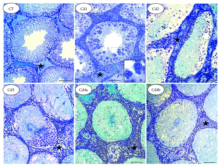
Figure 2.
Representative microscopic images of the tubular compartment in the testis from control rats and those exposed to cadmium (Cd). Control (CT): 0.9% saline; Cd1: 0.67 mg Cd/kg; Cd2: 0.74 mg Cd/kg; Cd3: 0.86 mg Cd/kg; and Cd4: 1.1 mg Cd/kg. In CT, well-defined tubular structure with the preserved seminiferous epithelium is observed. In Cd1 to Cd4, dose-dependent epithelial damage is observed, with intense germ cell dissociation and reduced distribution. In Cd1, germ cells with abnormal nuclear morphology are highlighted. Marked inflammatory infiltrate is observed in intertubular compartment (star), especially in Cd2 to Cd4.

