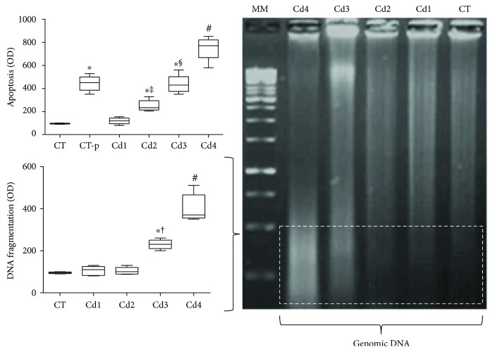
Figure 4.
Genomic DNA oxidation and apoptotic index in the testis from control rats and those exposed to cadmium (Cd). DNA oxidation was evaluated by a ladder test using electrophoresis in agarose gel (left image). Optical density was computationally evaluated from the third tercile of agarose gel (dotted area). Apoptosis was evaluated by using microscopic images obtained from testis histological sections submitted to the TUNEL technique. In the graphics, DNA oxidation and apoptosis results were expressed as the difference in optical density (OD) compared to those of negative control animals (CT). MM: molecular marker. Control (CT): 0.9% saline; CT-p: 1.00 U/mL DNase I (positive control for the TUNEL technique); Cd1: 0.67 mg Cd/kg; Cd2: 0.74 mg Cd/kg; Cd3: 0.86 mg Cd/kg; and Cd4: 1.1 mg Cd/kg. In the graphics, data are expressed as mean and standard deviation (mean ± SD). Statistical difference (∗p < 0.05 versus CT; ‡p < 0.05 versus CT-p and Cd1; §p < 0.05 versus CT-p, Cd1, and Cd2; #p < 0.05 versus CT, CT-p, Cd1, Cd2, and Cd3; and †p < 0.05 versus Cd1 and Cd2).

