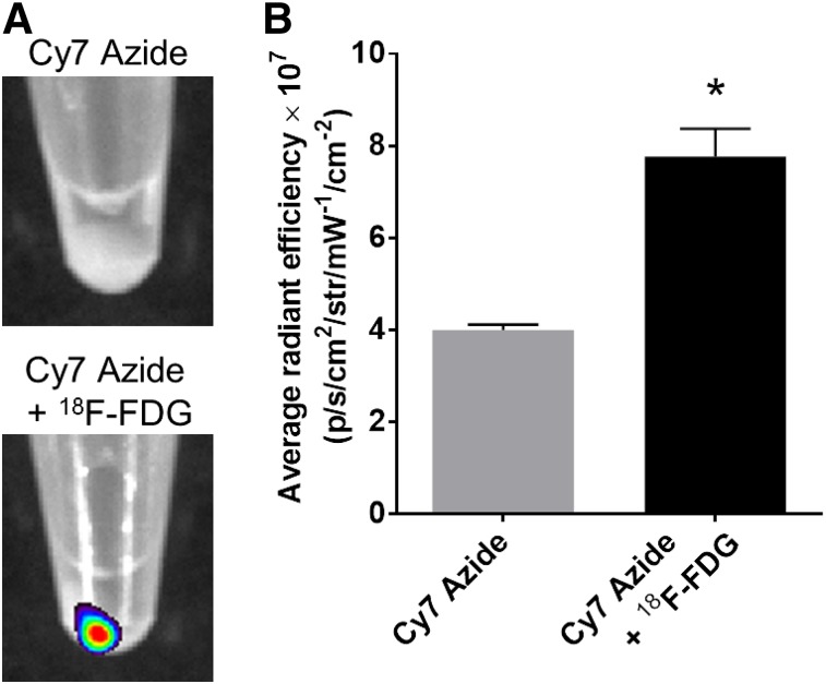
FIGURE 3.
CL-activated cell labeling using Cy7 azide. (A) Fluorescence images of HT1080 cell pellets incubated with 18F-FDG and Cy7 azide or Cy7 azide alone. (B) Quantitative analysis of HT1080 cell pellet fluorescence signal after incubation with Cy7 azide alone or together with 18F-FDG (P = 0.0004). IVIS Spectrum system was used for fluorescence imaging (emission filter, 800 nm; excitation filter, 745 nm; epiillumination; bins, 4; field of view, 24.4; f2, 0.75 s). *P < 0.05.

