Figure 1.
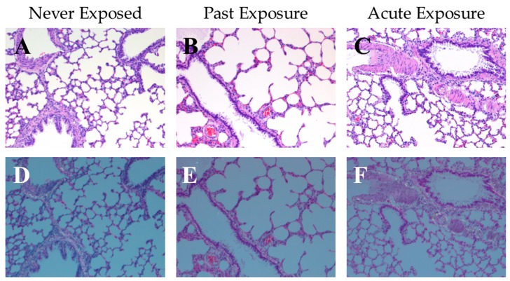
Representative photos of rat lung tissue at 100×; sections show an inflated area that included an arteriole, terminal bronchiole, and a larger bronchiole. (A–C) Hematoxylin and eosin (HE) stained tissue under bright light microscopy, and (D–F) corresponding HE stained tissue under polarized light. (A,D) tissue from a naïve rat that were never exposed to hydrophobic sand (“Never Exposed”); (B,E) tissue from a rat that underwent all LabSand experimental sessions, and was euthanized 1 month after the conclusion of the experiment with no further exposure to hydrophobic sand (“Past Exposure”); (C,F) tissue from a rat that underwent all LabSand experimental but was also placed in a cage with hydrophobic sand for 2 h prior to euthanasia (“Acute Exposure”).

