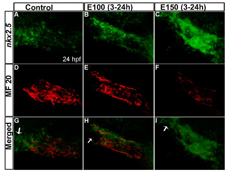
Figure 3.
Myocardial differentiation was delayed in ethanol-exposed embryos. (A–C) In situ hybridization detecting nkx2.5 expression showed weak nkx2.5 expression in the linear heart tube (indicated fewer undifferentiated FHF derived cells) but strong expression at the outflow pole (indicated arrival of second heart field (SHF) progenitors) in the control embryo (A); E100 embryos displayed strong nkx2.5 expression in the linear heart tube but no expression at the outflow pole (B); E150 embryos showed strong nkx2.5 expression in the linear heart tube and at the outflow pole (C); (D–F) Strong MF20 antibody staining in the linear heart tube in control embryos labeled differentiated cardiomyocytes (D); MF 20 staining was weaker in the heart tube in ethanol-treated embryos (E,F); (G–I) Co-labeled images showed myocardial differentiation delay in ethanol-exposed embryos. White arrows: pointing to the outflow pole.

