Figure 4.
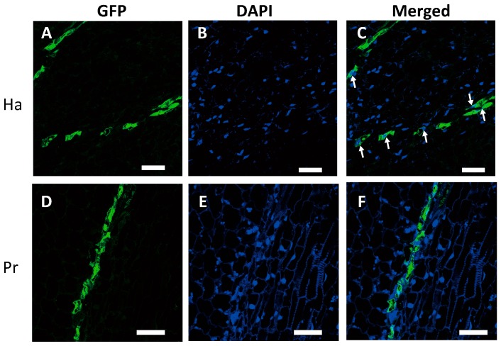
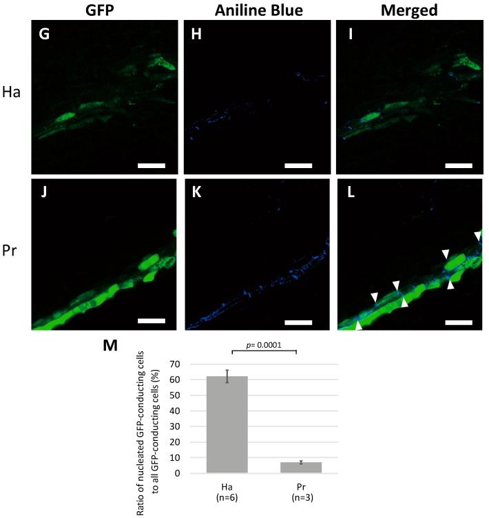
Detection of nuclei and callose-rich sieve plates in green fluorescent protein (GFP)-conducting cells in a 1-cm-diameter tubercle. (A–C) 4′6-diamidino-2-phenylindole (DAPI) staining of haustorial cells; (A) GFP, (B) DAPI, and (C) merged. (D–F) DAPI staining of protrusion cells; (D) GFP, (E) DAPI, and (F) merged. White arrows indicate nuclei. (G–I) Aniline Blue staining of haustorial cells; (G) GFP, (H) Aniline Blue, and (I) merged. (J–L) Aniline Blue staining of protrusion cells; (J) GFP, (H) Aniline Blue, and (I) merged. White triangles indicate sieve plates. Scale bar: 50 μm. (M) Percentage of nucleated GFP-conducting cells to all GFP-conducting cells. Ha, haustorium; Pr, protrusion. The means and standard deviations of six and three different haustorial and protrusion specimens, respectively, are presented. n: number of specimens.


